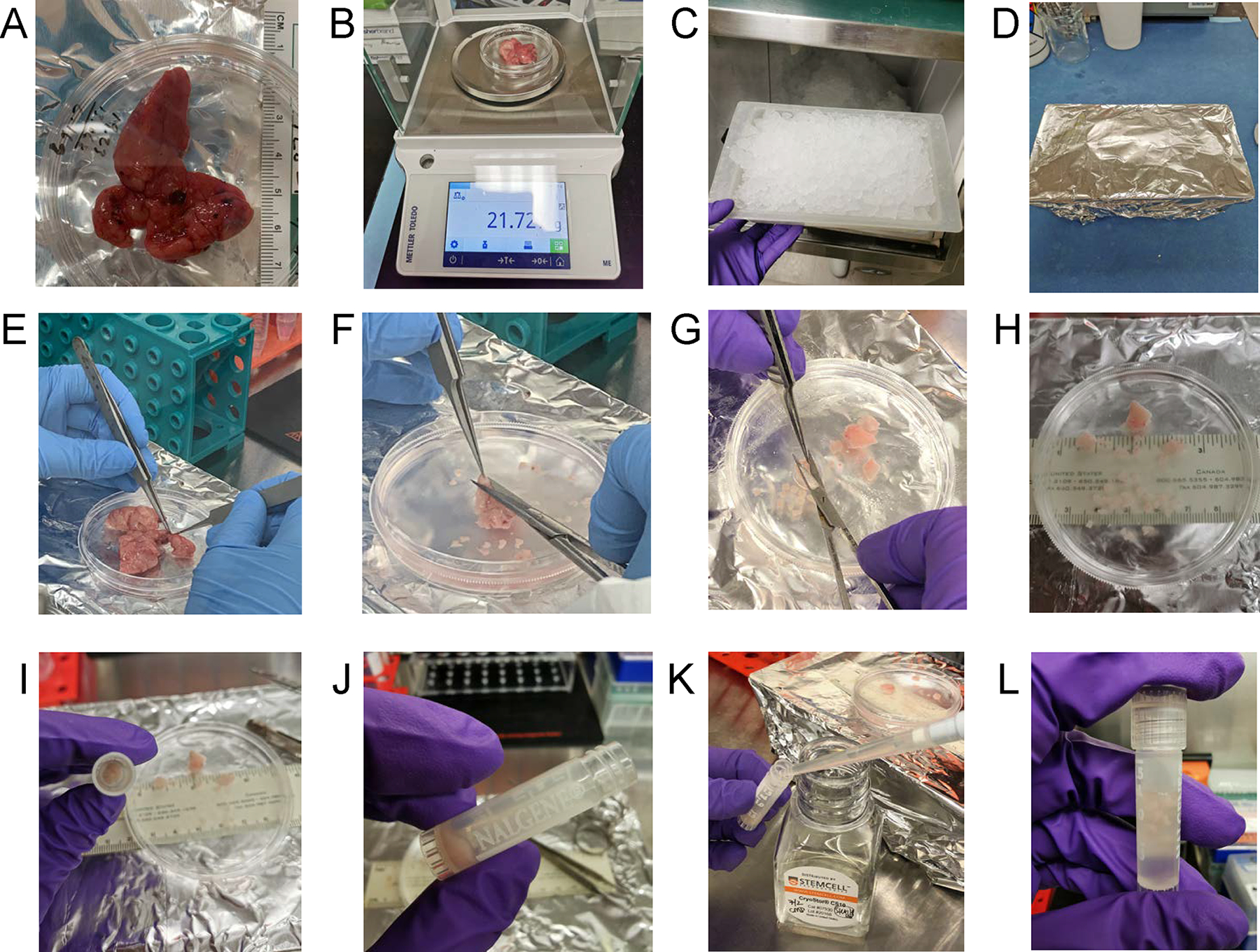
Figure 4.

Neonatal Thymus Processing. Using aseptic technique, the thymus tissue is (A) photographed and measured for length and (B) weighed. (C) A wet ice tray is prepared, (D) wrapped in aluminum foil, sprayed with 70% ethanol and placed in the biosafety cabinet. (E) The thymus tissue is placed on ice tray for dissection in cold PBS, whereupon blood vessels, adipose, and cauterized material is removed and then the lobes are cut into large sections. (F) Thymus is dissected into chunks approximately 1–3 cm × 1–3 cm. (G) The chunks are then cut into smaller pieces and moved to the side of the dish for (H) size determination and counting. Fragments are made into 1 mm × 1 mm size for cryopreservation in (I) cryotubes. Typically, 20 fragments per tube are frozen. (J) Fragments are settled by gravity and (K) 1ml of Cryostor CS10 freezing medium is added per vial. (L) The cap is tightened and each vial is inspected to ensure that fragments are submerged in liquid prior to being frozen to −80 degrees Celsius in a Corning CoolCell controlled-rate freezing container.
