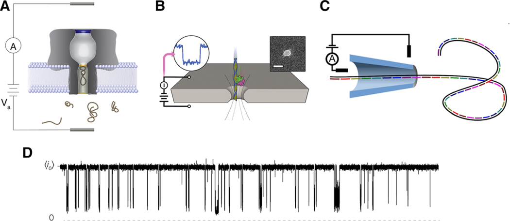
Figure 1.
Schematic illustrations of the different nanopore sensing configurations discussed herein. A) Biological pores are embedded in lipid bilayer membranes. B) Etched pores are nanoscale holes formed in solid-state supports such as silicon nitride or graphene. The length scales for these systems are typically on the order of 5–10 nm. C) Nanopore sensing has also been demonstrated with glass nanopipettes where length scales tend to be on the order of 50 nm. In all the three examples, target analyte enters into the sensing region of the pore and reduce the flow of current across the boundary. D) This gives rise to sizable current blockades whose depth (with respect to the open pore current ) and duration can inform about the physical and chemical attributes of the analyte under investigation. Data shown here is a 20 s current trace of 20 μm neurotensin interacting with αHL under a 70 mV applied transmembrane potential in 3 m KCl at pH 7.2. Images B and C are reproduced with permission.[98,101] Copyright 2014, ACS Publications and Copyright 2015, American Chemical Society.

