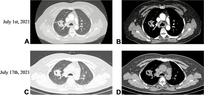
Fig. 1.
Chest CT at different time points. July 1st, 2021 Chest enhanced CT showed irregular soft-tissue density mass in the upper lobe of the right lung, and the bronchial branch of the upper lobe of the right lung was invaded and narrowed, which suggested a high possibility of lung cancer. There were also several small punctate calcification foci in the left lung (A). Slight calcification in mediastinum and left hilar lymph nodes (B). July 17th, 2021 After antifungal therapy, CT showed irregular soft tissue density shadow in the upper lobe of the right lung with cavity formation, which was considered to be lung cancer, with little change from the CT result before treatment (C, D)

