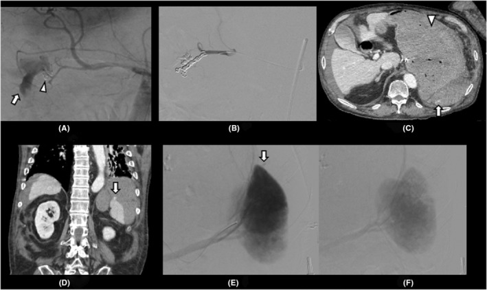
FIGURE 2.

(A) Angiography shows extravasation from the right gastroepiploic artery and anterior superior pancreaticoduodenal artery into the duodenal lumen (arrow). Clips used for trying to stop bleeding are shown (arrowhead). (B) Metal coils are placed from the gastroduodenal artery to these two branches for embolization, and no extravasation is observed. (C, D) Contrast‐enhanced abdominal computed tomography on the second day shows seudoaneurysm from the spleen (arrow) and a large volume of contents but no extravasation in the stomach (arrowhead). (E) Angiography shows evidence of pseudoaneurysm from the upper pole of the spleen (arrow). (F) After embolization of the splenic artery with a gelatin sponge, no pseudoaneurysm is observed.
