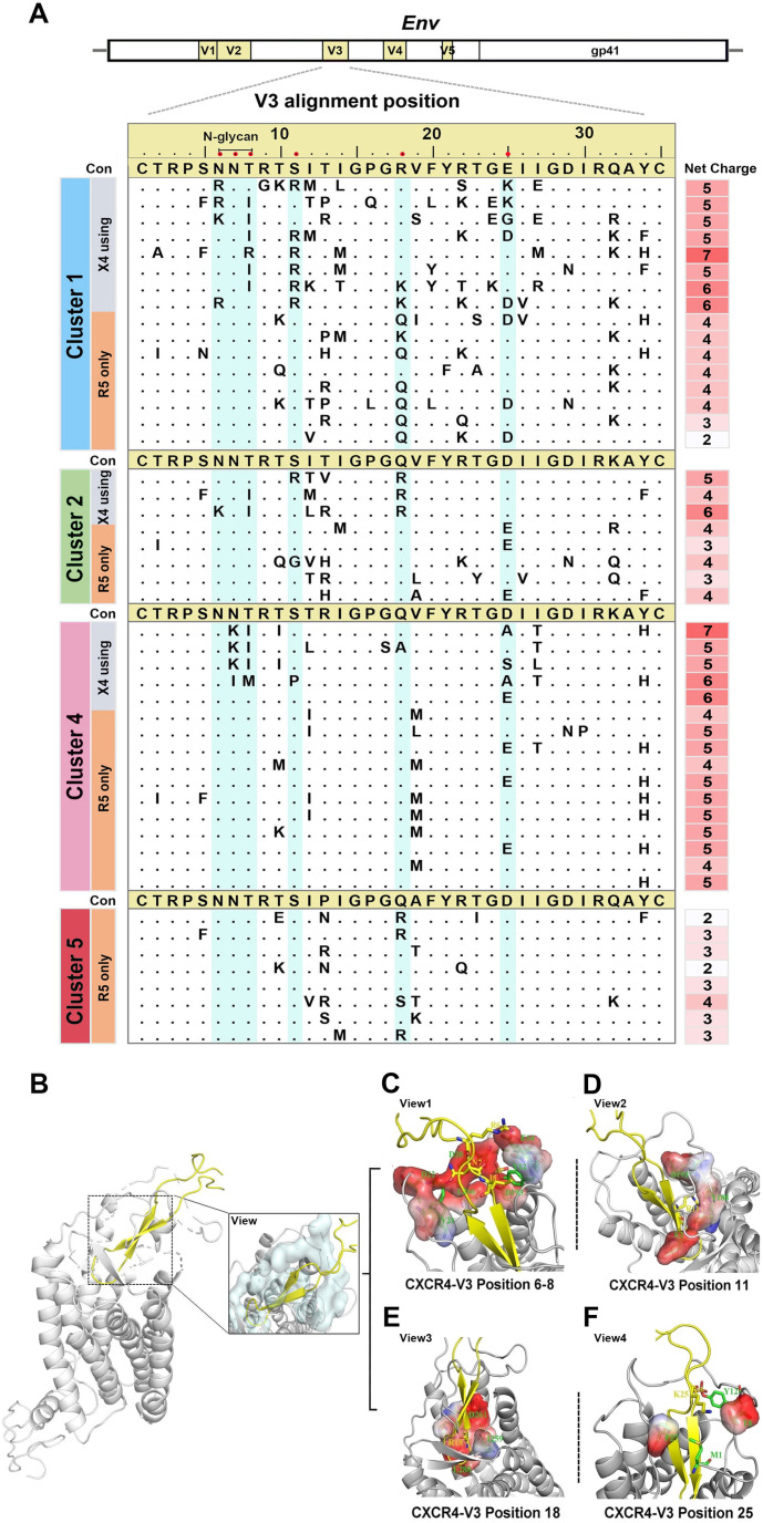
Fig 4.
Genetic features of CXCR4 sequences with phenotypic confirmation and structural analyses of co-receptor binding models. (A) Amino acid positions and net charge distribution in the V3 region of phenotype-confirmed primary viral isolates with CXCR4 usage. Crucial CXCR4 utilization-associated V3 positions are highlighted in cyan. (B) Structural modeling of the binding site between the co-receptor CXCR4 and the V3 region using the docking model. The interfaces between co-receptor CXCR4 and V3 are depicted with an overall side view and detailed in a close-up view (right boxed). (C–F) Detailed description of the interactions with the major sites of the V3 loop in the CXCR4 binding pocket. The surface of CXCR4 binding to V3 is displayed with negative (red colored) and positive (blue colored) electrostatic potentials. Key residues are depicted as sticks, and the overall structure is shown as a graphical representation.

