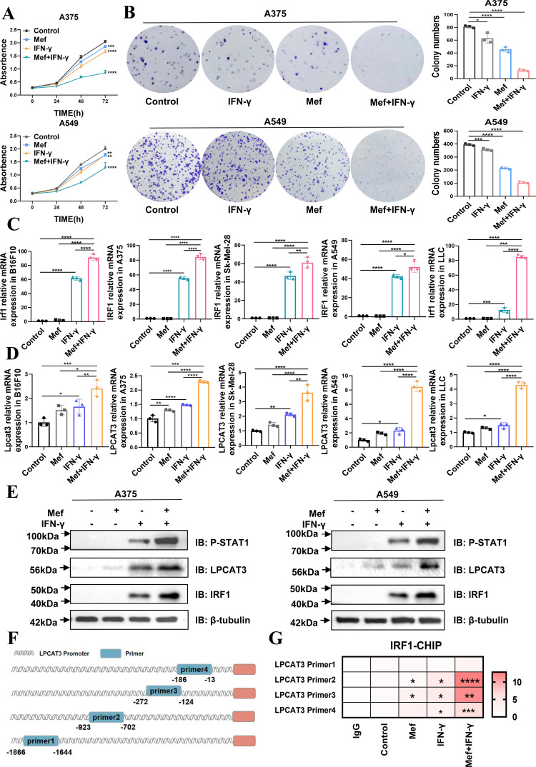
Figure 3.
Mefloquine in combination with IFN-γ inhibits tumor growth through the activation of Lpcat3. (A) The numbers of A375 and A549 cells in cultures supplemented with Mef (4 µM) or IFN-γ (20 ng/mL) for 24, 48, or 72 hours were determined (n=6 replicate cultures for each cell type). (B) A colony formation assay of A375 and A549 cells in cultures supplemented with Mef (4 µM) or IFN-γ (20 ng/mL) for 48 hours (n=3 replicate cultures for each cell type). (C–D) The relative mRNA expression of LPCAT3 and IRF1 in A375 and A549 cells treated for 48 hours with Mef (4 µM) or IFN-γ (20 ng/mL) was determined (n=3 replicate cultures for each cell type). (E) Immunoblots of LPCAT3, IRF1, and P-STAT1 in A375 and A549 cells after Mef (4 µM) or IFN-γ (20 ng/mL) treatment for 6 hours are shown. (F) Primers were designed for the LPCAT3 promoter region based on JASPAR predictions. (G) IRF1 binding to the promoter of LPCAT3 in A375 cells was validated by ChIP-qPCR (n=3 replicate cultures for each cell type). All the data are presented as the mean (±SD). One-way analysis of variance was performed for comparisons among multiple groups. Statistical significance was assessed according to statistical methods (*p<0.05; **p<0.01; and ***p<0.001). ChIP, chromatin immunoprecipitation; IFN, interferon; LLC, Lewis lung cancer; LPCAT3, lysophosphatidylcholine acyltransferase 3; Mef, mefloquine; mRNA, messenger RNA; qPCR, Quantitative Real-time PCR.

