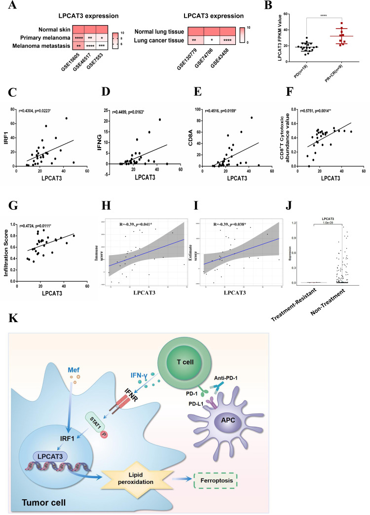
Figure 7.
LPCAT3 is associated with an improved immune microenvironment and immunotherapeutic efficacy. (A) Data from the GEO database were used to assess LPCAT3 expression in patient-derived primary melanoma, metastatic melanoma, normal skin, lung cancer tissue and normal lung tissue. (B) Data from the GEO database (GSE 91061 (PD (n=19), PR+CR (n=9))) were used to assess LPCAT3 expression in patients with anti-PD-1-treated melanoma. (C–E) Data from the GEO database (GSE91061) was used to analyze the correlation between LPCAT3 and IRF1 (C), IFN-γ (D) and CD8a (E) after anti-PD-1 therapy. (F–G) ImmuCellAI was used to analyze the correlation between LPCAT3 expression and CD8+ T-cell enrichment (F) and LPCAT3 expression and immune cell infiltration (G) in the immune microenvironment. (H–I) The ESTIMATE algorithm was used to analyze the correlation between LPCAT3 expression and immune score (H) and LPCAT3 expression and ESTIMATE score (I) in PD and PR CR patients from the GSE91061 data set. (J) Single-cell sequencing was used to analyze the expression of LPCAT3 in melanoma cells resistant to anti-PD-1 immunotherapy. (K) A diagram of the molecular mechanism described in the article is shown. All the data are presented as the mean (±SD). A t-test was used for comparisons between two groups. One-way analysis of variance was performed for comparisons among multiple groups. Pearson correlation coefficient was performed for correlation analysis. Statistical significance was assessed according to statistical methods (*p<0.05; **p<0.01; and ***p<0.001). CR, complete response; GEO, Gene Expression Omnibus; IFN, interferon; LPCAT3, lysophosphatidylcholine acyltransferase 3; Mef, mefloquine; PR, partial response; PD, progressive disease; PD-1, programmed cell death 1; PD-L1, programmed death ligand 1.

