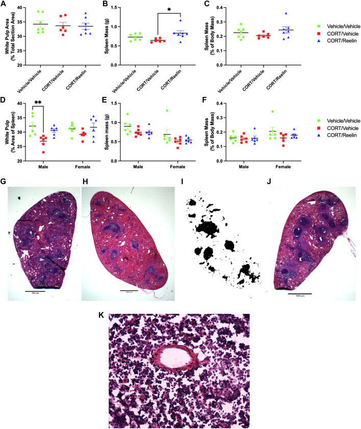
FIGURE 6.
Analysis of spleens shows that Reelin elevated spleen mass in females exposed to 1.3 cycles, and CORT reduced the white pulp area in males only, which was partially recovered with Reelin treatment. (A) Percentage of the spleen surface area occupied by white pulp in rats exposed to 1.3 cycles of chronic stress, showing no change in the white pulp percent area. (B) Spleen mass of rats exposed to 1.3 cycles of chronic stress, showing no significant deficit in spleen mass with chronic stress, but animals exposed to both chronic stress and Reelin injections had heavier spleens than rats exposed to chronic stress alone. (C) Spleen mass expressed as the percentage of body mass, showing no significant differences in rats exposed to 1.3 cycles of chronic stress. (D) Percentage of the spleen surface area occupied by white pulp in rats exposed to 2.6 cycles of chronic stress, showing a deficit in the white pulp area in males but not females, which was partially restored after Reelin treatment. (E) Spleen mass in animals exposed to 2.6 cycles of chronic stress treatment, showing no significant difference in the mass between treatments. (F) Spleen mass expressed as the percentage of total body mass for animals exposed to 2.6 cycles of chronic stress treatment, showing no significant change in the spleen mass relative to body mass between any treatment groups. (G) Photomicrograph at ×1 magnification demonstrating the H&E staining of a section obtained from a male rat exposed to vehicle/vehicle injections. (H) Photomicrograph at ×1 magnification of the spleen from a CORT/vehicle-treated male rat after H&E staining. (I) Binarization of Image 8H demonstrating how H&E-stained images were converted to black and white for quantifying white pulp. (J) Photomicrograph at ×1 magnification of the spleen from a CORT/Reelin-treated male rat after H&E staining. (K) Photomicrograph at ×40 magnification of an H&E-stained spleen section within the white pulp region from a vehicle-treated rat exposed to 2.6 cycles of injections that shows the presence of artifacts, likely created during cryosectioning.

