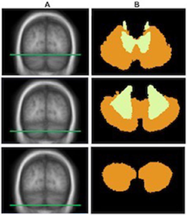
Figure 1.
Probabilistic cerebellar labelling scheme of the FreeSurfer atlas. Cerebellar segmentation into cortical versus subcortical components using the probabilistic FreeSurfer atlas. Three distinct axial slices of the cerebellum as labelled in the probabilistic FreeSurfer atlas in the standardised MNI305 brain. For each axial slice through the T1-weighted image (A), the corresponding parcellation of the cerebellum (B) into cortical (orange) and subcortical (yellow) regions is shown.

