Figure 1.

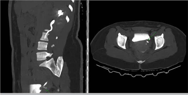
Computed tomography with intravenous contrast demonstrates a fistula extending from the distal left ureter to the vagina.

Computed tomography with intravenous contrast demonstrates a fistula extending from the distal left ureter to the vagina.