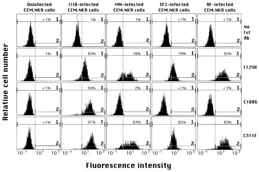
FIG. 1.
Results of flow cytometry analyses of uninfected or HIV-1-infected CEM.NKR cells. MAbs 1125H, C108G, and C311E were used at 20 μg/ml. In this experiment, gate 2 was set to exclude ≥99% of the background fluorescence observed with no first Ab added (only fluorescein isothiocyanate conjugate was added) for each type of infected or uninfected cells. The percentage of cells staining within gate 2 (above background) is shown in each panel.

