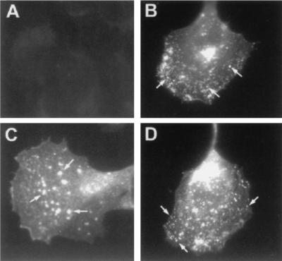
FIG. 9.
Immunofluorescence staining with anti-P37 antibody. CV-1 cells were either mock infected (A) or infected with WR (B), W-B5R− (C), or W-B5RΔSCR1–4 (D). At 7 h postinfection, cells were fixed, made permeable, washed, and then incubated with a monoclonal antibody to P37, the major protein present in EEV. To visualize the primary antibody, rabbit anti-rat antibody coupled to FITC was used. Representative cells were photographed. Arrows indicate small punctate staining of WR and W-B5RΔSCR1–4 and large fluorescent dots in the B5R deletion virus (C).

