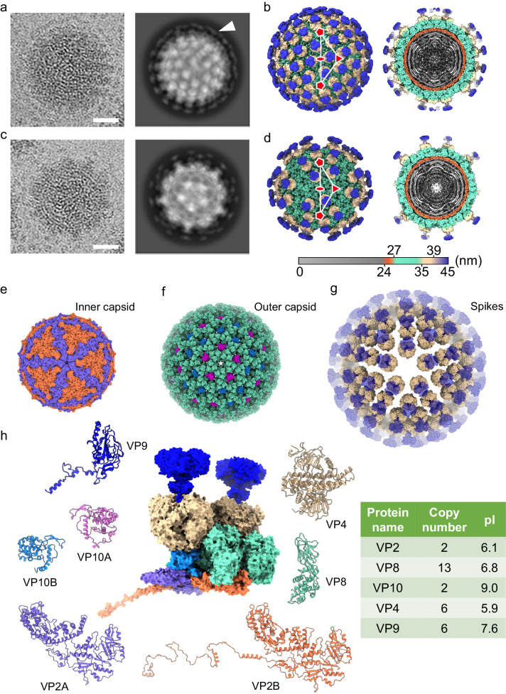
Fig. 1. Structural overview of full and partial Banna virions (BAVs).
a Representative 2D image (left) and class averaging (right) of BAV full particles. The spiky density on the viral surface forms a broken ring indicated by an arrow. Scale bar: 20 nm. b Surface representation of the full BAV particle viewed along a twofold axis (left) and a cutaway view showing the internal density of the virus (right). The virus is radially colored, showing the viral genome and RdRp (gray), inner capsid shell (orange), outer capsid shell (emerald), and spikes (beige and blue). An asymmetric unit (ASU) is marked by a white triangle. One twofold, one threefold, and two fivefold axes are indicated by an ellipse, a triangle, and two pentagons, respectively. c 2D image (left) and class averaging (right) of BAV partial particles. d Surface representation (left) and cutaway view (right) of BAV partial particle. e–g Surface representation of inner capsid shell (e), outer capsid shell (f), and spikes (g) of full BAV particles viewed along a fivefold axis. Maps are presented as solvent-excluded surfaces with a probe radius of 1.4 Å. Proteins are colored as in (h). Density for VP9 is shown as a transparent blue surface. h Protein composition of an ASU (center, presented as a solvent-excluded surface) of full BAV particles and ribbon diagrams of individual proteins. Copy number and pI values are listed in the table (right).

