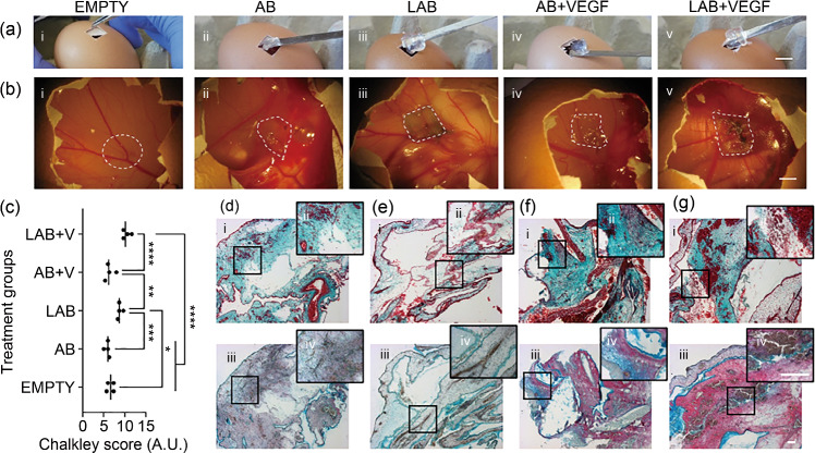
Fig. 6.
Nanoclay-based inks support sustained release of VEGF in the CAM model. Macrographs during a sample implantation and b retrieval: (i) empty, (ii) AB, (iii) LAB, and VEGF-loaded (iv) AB and (v) LAB 3D-printed scaffolds. c Chalkley score of vascularized samples and controls. d–g Histological micrographs of samples stained for (i, ii) Goldner’s Trichrome and (iii, iv) Alcian Blue & Sirius Red. Statistical significance was assessed using one-way ANOVA. Data are presented as mean±standard deviation, n=4, *p<0.05, **p<0.01, ***p<0.001, ****p<0.0001. Scale bars: a, b 10 mm, d–g 100 µm. VEGF: vascular endothelial growth factor; CAM: chick chorioallantoic membrane; AB: alginate-bone-ECM; LAB: Laponite-alginate-bone-ECM; ANOVA: analysis of variance

