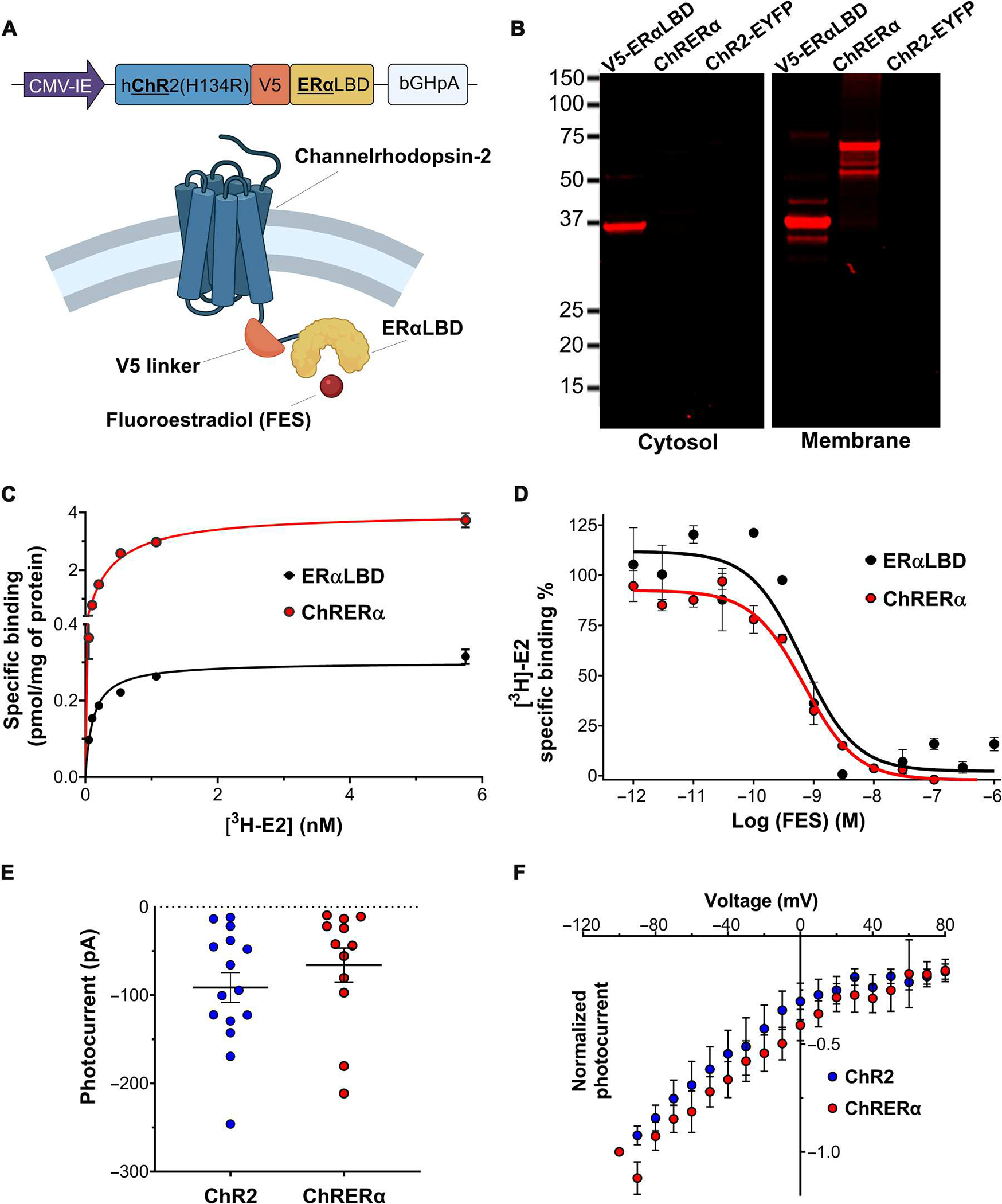
Fig. 1. Development and characterization of ChRERα in HEK-293 cells.

(A) Schematic illustrations of the encoding sequence for ChRERα (top) and the protein structure of ChRERα (bottom) are shown. (B) Representative Western blots verifying the subcellular localization of the V5 epitope in cytosolic or membrane fractions of HEK-293 cells transfected with V5-ERα-LBD, ChRERα, or ChR2-EYFP are shown. (C) [3H]E2 binding saturation curves in membrane homogenates from HEK-293 cells transfected with ERα-LBD (black) or ChRERα (red). (D) [3H]E2 competition binding curves with FES in membrane homogenates from HEK-293 cells transfected with ERα-LBD or ChRERα. Values for fitted parameters are described in the main text. (E) Photocurrent amplitudes in picoamperes (pA) and (F) light-induced voltage-current curves in millivolts (mV) of HEK-293 cells transfected with ChR2 (blue) or ChRERα (red). All data are shown as means ± SEM except that dots in (E) are individual cells.
