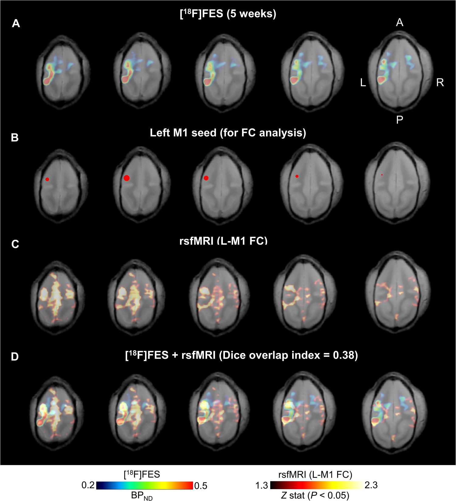
Fig. 7. [18F]FES-PET and ChRERα correlate with functional brain connectivity in NHPs.

(A) Horizontal sections (left, most dorsal; right, most ventral) of [18F]FES-PET from squirrel monkey 1 at 5 weeks after AAV. (B) Left M1 seed (red 2-mm sphere centered at peak [18F]FES signal) for functional connectivity analysis of resting state functional MRI (rsfMRI). (C) rsfMRI functional connectivity patterns of the left M1 seed in an independent group of squirrel monkeys (n = 9, 35 total scans) coregistered to the squirrel monkey 1 structural MRI. (D) Overlapping patterns of [18F]FES binding (ChRERα expression) and rsfMRI suggest structural and functional connectivity between left M1 and ipsilateral PPC and in the contralateral hemisphere (r = 0.23; P = 0 with Fisher’s z transformation; 27,871 voxels; and Dice overlap index = 0.38).
