. 2024 Mar 14;14(13):8718–8725. doi: 10.1039/d4ra01617c

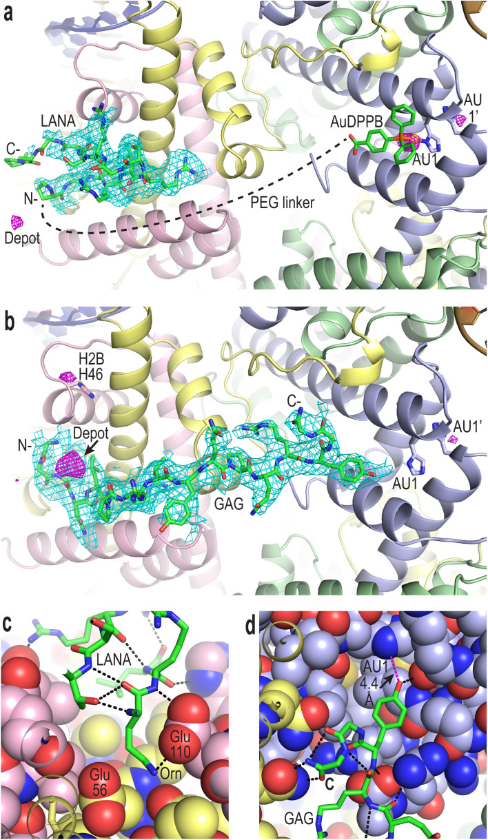
Fig. 2. Viral peptide binding and AuDPPBX coordination/localization in the nucleosome. (a and b) X-ray crystal structure of NCP treated with Au–LANA ((a); 2 N- and 5 C-terminal peptide residues not shown/visible in electron density maps) and Au–GAG (b). A FOFC omit electron density map (cyan; contoured at 2σ; prior to inclusion of viral peptide) and an anomalous difference electron density map (magenta; contoured at 3.5σ; indicates locations of gold atoms) are shown superimposed on the model (N- and C-, peptide N- and C-termini). (c and d) Close view of the LANA (c) and GAG (d) conjugate peptides binding in the NCP crystal structures (H-bonds, dashed black lines; close contact, dashed magenta line; histone protein atoms in space filling representation). (a–d) Histone proteins are shown in purple (H3), pale green (H4), yellow (H2A) and pink (H2B), and DNA strands (a and b) are orange and dark blue.

Fig. 2