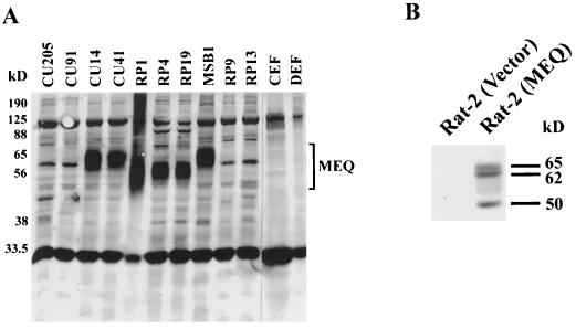
FIG. 1.
Constitutive expression of MEQ proteins in the MDV tumor cell lines and MEQ-transformed Rat-2 cells. Total cell lysates were prepared from MDV cell lines (RP1, RP4, RP19, and MSB1), MDV-infected T-cell lines (CU14 and CU41), reticuloendotheliosis virus cell lines (CU91, CU205, and RP13), avian leukosis virus cell line (RP9), and normal avian embryo fibroblasts (CEF and DEF) (A) as well as from MEQ-transformed and vector-infected Rat-2 cells (B). Cell lysates equivalent to approximately 0.5 × 10⁶ to 1 × 10⁶ cells per lane were analyzed by SDS–10% PAGE. The gels were transferred to Immobilon polyvinylidene difluoride membranes and blotted with MEQ antisera (1:4,000 dilution). The Western blots were subsequently detected with the conventional alkaline phosphatase method.

