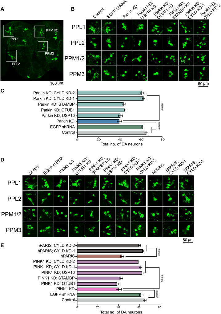
Fig. 2. Validation of candidate DUBs identified in primary and secondary RNAi screens places Drosophila cylindromatosis (dCYLD) in the PINK1/parkin pathway.
(A) Representative confocal image of the Drosophila whole brain showing the PPL1, PPL2, PPM1/2, and PPM3 DA neuron clusters. Scale bar, 100 μM. (B) Representative confocal images of individual dopamine neuron clusters visualized using TH immunofluorescence in the indicated genotypes under conditions of parkin KD. Scale bar, 50 μM. N = 10 flies per genotype. (C) Quantification of neuronal numbers within individual dopamine neuron clusters in the indicated genotypes. (D) Representative confocal images of individual dopamine neuron clusters visualized using TH immunofluorescence in the indicated genotypes under conditions of PINK1 KD or hPARIS overexpression. Scale bar, 50 μM. (E) Summary of dopamine neuron quantifications in the indicated genotypes. TH-Gal4/+ flies served as control. N = 10 flies per genotype. Quantitative data = means ± SEM. One-way ANOVA; ****P < 0.0001. See also fig. S3.

