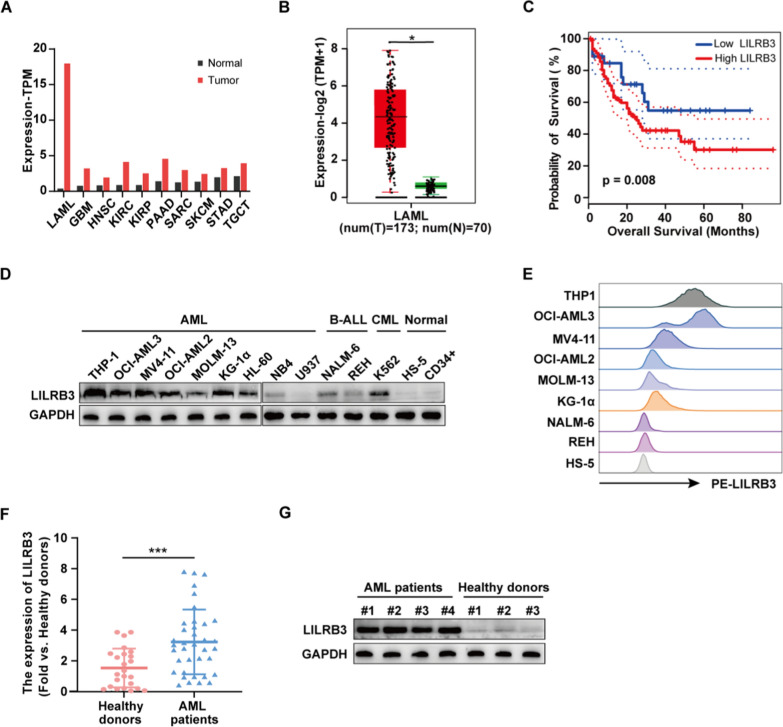
Fig. 1.
LILRB3 is highly expressed in patients with AML and AML cells. A The expression profile of LILRB3 across TCGA datasets. Images were obtained from the GEPIA online database (http://gepia2.cancer-pku.cn/). LAML acute myeloid leukemia, GBM:glioblastoma multiforme, HNSC head and neck squamous cell carcinoma, KIRC kidney renal clear cell carcinoma, KIRP kidney renal papillary cell carcinoma, PAAD pancreatic adenocarcinoma, SARC sarcoma, SKCM skin cutaneous Melanoma, STAD stomach adenocarcinoma, TGCT testicular germ cell tumors. B Expression levels of LILRB3 in AML from TCGA datasets. Tumor: acute myeloid leukemia; normal: normal donors. C Kaplan–Meier overall survival curves for 173 AML patients classified according to relative LILRB3 expression level. Patients with higher LILRB3 had a shorter survival. D The LILRB3 protein level in a panel of malignant hematological cells, normal bone marrow cell lines (HS-5), and peripheral blood CD34 + cells from healthy donors. E LILRB3 expression on the AML cell surface was higher than that on B-ALL cell lines and HS-5. F–G LILRB3 mRNA and protein levels in BMMCs of AML patients and healthy donors in our clinical samples. *P < 0.05, **P < 0.01, ***P < 0.001

