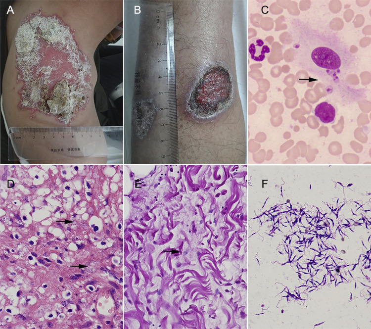
Fig 3. Dermatological, pathological and etiological features of CL skin lesion.
A. The representative picture of nodular lesion (dry type). B. ulcerative lesion (wet type). C. Leishmania amastigotes detected in lesion smear with Giemsa staining (1000×, arrow). D-E. Pathological examination of skin lesion biopsy showing amastigotes (arrowed) stained with H&E (D) or PSA (E) (400×). F. Leishmania promastigotes detected in lesion scraping culture (Giemsa staining, 1000×).

