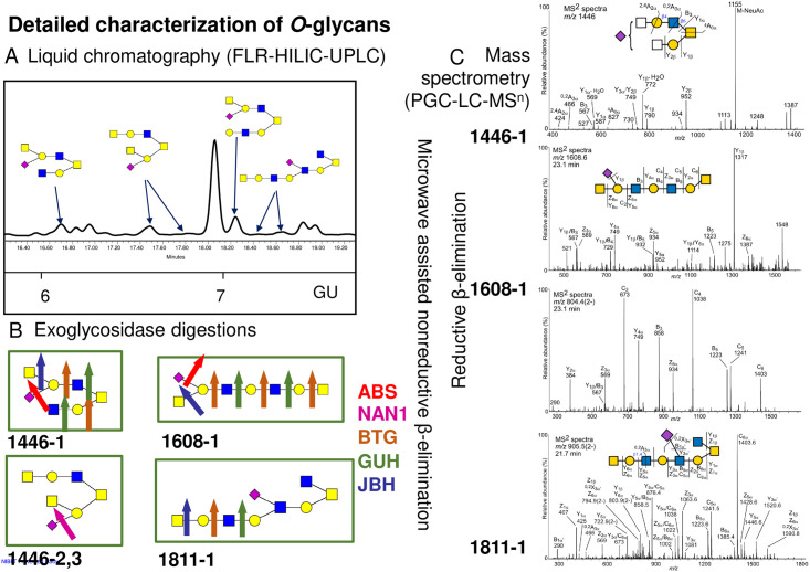
Fig 3. Novel glycans found in ileal samples and their detailed characterization using combination of FLR-HILIC-UPLC, exoglycosidase digestions and PGC-LC-MSn.
A) Part of HILIC-UPLC chromatogram with novel glycans pictured. B) Novel glycans digested by exoglycosidase enzymes, namely α2-3/6/8 sialidase (ABS), α2-3/8 sialidase (NAN1), β1-3/4/6 galactosidase (BTG), β1-2/4/6 hexosamindase (GUH) and β1-2/3/4/6 hexosamindase (JBH). Detailed assignments are in S3B Table. C) 1446. MS/MS spectra of a reduced and nonderivatized sialylated O-glycan ‘1446–1’ (S2B Table) eluting at 20.6 min and detected at m/z 1446.5 ([M-H]- precursor ion) in ileal mucus of WT control mice. 1608. MS/MS spectra of the mono- and doubly charged precursor ions of a reduced and nonderivatized sialylated O-glycan ‘1608–1’ (S2B Table) eluting at 23.1 min and purified from ileal mucus of Cybanmf333 (Cyba mut) mice. Top panel: m/z 1608.6 ([M-H]- precursor ion); Bottom panel: m/z 804.4 ([M-2H]2- precursor ion). 1811. MS/MS spectra of a reduced and nonderivatized sialylated O-glycan ‘1811–1’ (S2B Table) eluting at 21.7 min and detected at m/z 905.5 ([M-2H]2- precursor ion) in ileal mucus of WT control mice. The analyses were performed using porous graphitized carbon liquid chromatography and low-resolution mass spectrometry kept in the negative ion mode. Proposed structures are shown using SNFG [28] and glycan fragmentation is as defined by Domon and Costello [29].

