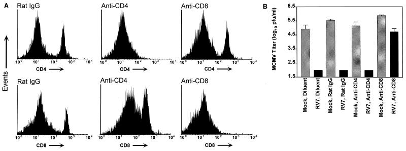
FIG. 6.
Effect of depletion of CD4 and CD8 T cells on RV7 vaccination. BALB/c mice were vaccinated with 2 × 10⁵ PFU of RV7 s.c., or were mock vaccinated, on days 0 and 14 of the experiment and then challenged with 104 PFU of sgMCMV i.p. on day 28 of two independent experiments. To deplete CD4 and CD8 T cells, rat monoclonal antibodies were passively administered to mice before and after challenge on day 28, using the doses and schedules specified in Materials and Methods. Purified rat IgG and the diluent used for preparing antibodies for injection were used as controls. Four days after challenge, spleens were harvested; one half was taken for determination of MCMV titer, and one half was used to determine the efficacy of CD4 and CD8 T-cell depletion by FACS analysis. (A) Single-color FACS histograms showing the proportions of CD4 and CD8 T cells in spleens from MCMV-infected mice after treatment with the indicated antibodies. Spleen cells from diluent-treated mice and rat IgG-treated mice were similar, and thus only FACS profiles from rat IgG-treated mice are shown. Two experiments were performed with different depletion regimens, and the FACS data shown are from the regimen using higher doses of anti-CD4 and anti-CD8 antibodies (see Materials and Methods). (B) MCMV titer in spleens of mice from the indicated groups. For CD8 depletion, and CD4 depletion in mock-vaccinated animals, data from two experiments using the different depletion regimens were pooled (n = 7 mice per condition). CD4 depletion in RV7-vaccinated mice using the lower-dose protocol resulted in only 57% depletion, so data are presented only from the experimental protocol using higher doses of anti-CD4 antibodies, which resulted in >99% depletion (n = 3 mice). Using the lower-dose protocol, where CD4 depletion was only 57%, we also failed to detect infectious MCMV in four of four RV7-vaccinated mice (not shown). When spleen titer was not detected by plaque assay, the titer was arbitrarily fixed at 100, the limit of plaque assay sensitivity. Data are shown as mean log titer ± SEM.

