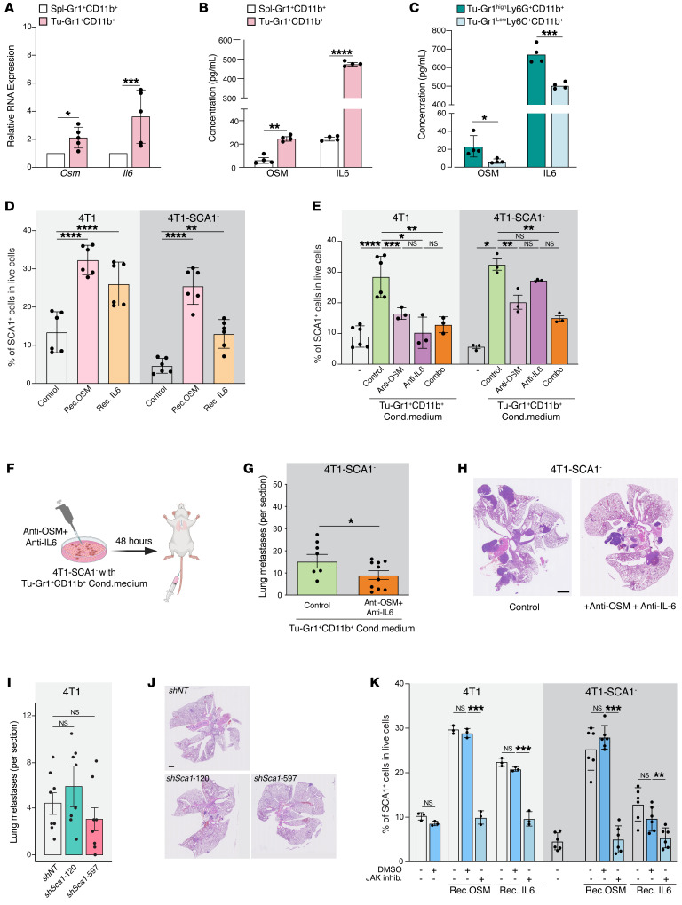
Figure 5. SCA1+ population is modulated by OSM/IL-6/JAK pathway.
(A) Relative Osm and Il6 mRNA expression in Tu-Gr1+CD11b+ and Spl-Gr1+CD11b+ cells. n = 4–5/group. (B and C) OSM and IL-6 protein quantification in supernatant of (B) Tu-Gr1+CD11b+ and Spl-Gr1+CD11b+ (C) and Tu-Gr1hiLy6G+CD11b+ and Gr1loLy6G–CD11b+ cells. n = 4/group. (D) Fraction of SCA1+ cells of parental 4T1 or sorted 4T1-SCA1– cells upon exposure to OSM or IL-6 (10 ng/ml 48 hours). n = 6/group. (E) Effect of inhibition of OSM and IL-6 in Tu-Gr1+CD11b+ conditioned medium on SCA1 expression in parental 4T1 or sorted 4T1-SCA1– cells after 48 hours treatment. n = 3–6/group. (F) Experimental design for examining lung metastatic capacity of IL-6/OSM. 4T1-SCA1– cells were primed by Tu-Gr1+CD11b+ conditioned medium with dual depletion of OSM/IL-6 depleted or control, in vitro for 48 hours and then injected into tail vein. Lungs were examined for metastasis 10 days later. (G and H) Quantification of lung metastases (G) and representative H&E staining images of lung sections (H). n = 8–10/group. Scale bar: 1 mm. (I and J) Quantification of lung metastases in mice injected with 4T1 control and 4T1 silenced for Sca1 (shSca1-120 and shSca1-597) (I). n = 8/group. Representative H&E stained lung sections (J). Scale bar: 1 mm. (K) SCA1+ population stimulation in cultured parental 4T1 or sorted 4T1-SCA1– cells by recombinant OSM or IL-6 (10 ng/ml, 48 hours) in vitro in presence of ruxolitinib (5 μM) or DMSO control. n = 3–6/group. Data are represented as means ± SEM from 3 independent experiments. P values were calculated using unpaired 2-tailed Student’s t test (A–C and G); unpaired 2-tailed Student’s t test with Holm’s correction (I); 1-way ANOVA with Dunnett’s multiple-comparison test (D and K); or 1-way ANOVA with Tukey’s multiple-comparison test (E). *P < 0.05; **P < 0.01; ***P < 0.001; ****P < 0.0001.

