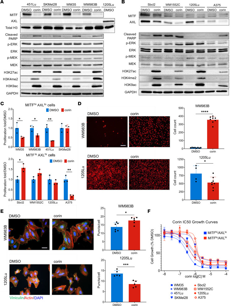
Figure 1. The CoREST repressor complex mediates phenotype switching in melanoma cell lines.
(A and B) Western blot analysis of the MITFhi/AXLlo melanoma cell lines 451Lu, SKMel28, WM35, and WM983B (A) and the MITFlo/AXLhi melanoma cell lines Sbcl2, WM1552C, 1205Lu, and A375 (B) following 24 hours of treatment with DMSO or 2.5 μM corin. 1205Lu lysates were used as a positive control for AXL in A. Western blots were run contemporaneously. (C) Cellular proliferation of MITFhi/AXLlo (upper panel) and MITFlo/AXLhi (lower panel) melanoma cell lines following 24 hours of treatment with DMSO or 2.5 μM corin (n = 3). (D) Invasion assay and quantification of MITFhi/AXLlo (WM983B, upper panel) and MITFlo/AXLhi (1205Lu, lower panel) melanoma cells following 24 hours treatment with DMSO or 2.5 μM corin (n = 4–8). Representative images are shown. Scale bar: 100 μm. (E) Vinculin staining of focal adhesions in MITFhi/AXLlo melanoma cells (WM983B, upper panel) and MITFlo/AXLhi melanoma cells (1205Lu, lower panel) following a 24-hour treatment with DMSO or 2.5 μM corin (n = 5–6), with quantification of focal adhesions (puncta/cell) on the right (n = 5–6). Representative images are shown. Scale bar: 20 μm. (F) Dose-response proliferation assays of MITFhi/AXLlo (WM35, WM983B, 451Lu, SkMel28) and MITFlo/AXLhi (Sbcl2, WM1552C, 1205Lu, A375) melanoma cell lines treated with increasing doses of corin for 72 hours. *P < 0.05, **P < 0.01, ***P < 0.001, and ****P < 0.0001, by 2-tailed, unpaired t test compared with DMSO controls.

