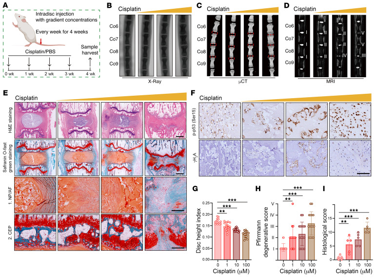
Figure 2. Genomic DNA damage–induced NP cell senescence promotes the degeneration of IVDs in vivo.
(A) Schematic illustration of the experimental design. (B–D) Representative x-ray images (B), μCT images (C), and MRI (D) of rat coccygeal IVDs treated with intradisc injection of cisplatin at different concentrations (n = 5). (E) H&E staining and SO&FG staining of rat coccygeal IVDs. Scale bar: 1 mm. (F) IHC staining of p-p53 and γH2A in rat coccygeal IVDs. Scale bar: 250 μm. (G–I) Disc height index (G) (n = 15 biological replicates), Pfirrmann degenerative grades (H) (n = 15 biological replicates) and histological score (I) (n = 5 biological replicates) of rat coccygeal IVDs. Data are presented as the mean ± SEM. At least 3 independent experiments were performed. **P < 0.01 and ***P < 0.001, by 2-way ANOVA (G–I).

