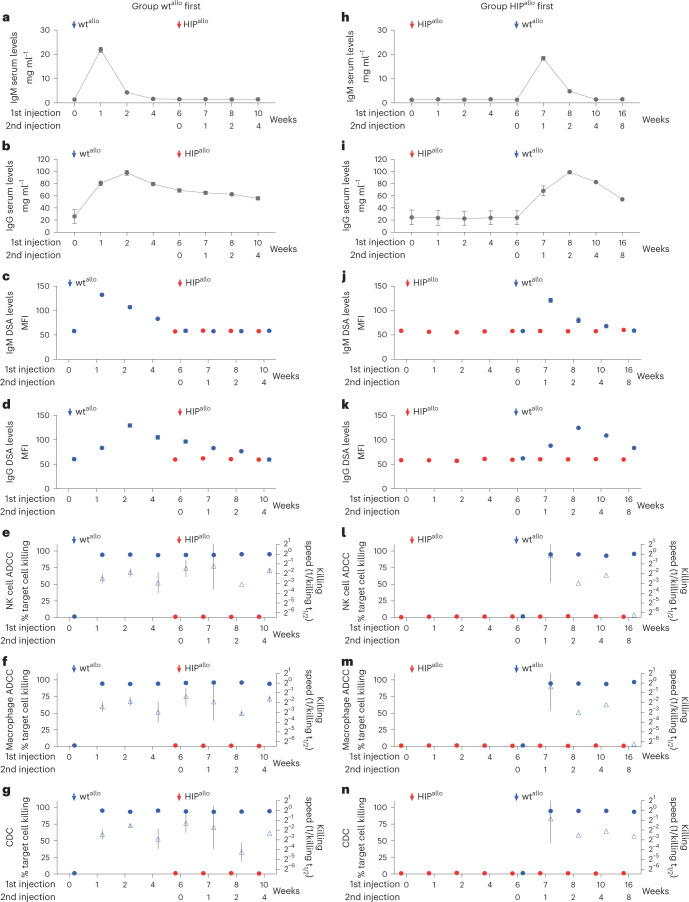
Fig. 2. Antibody-mediated responses against allogeneic rhesus macaque wt and HIP grafts.
a–g, Antibody immune assays in the group receiving wtallo first. Total serum IgM (a) and IgG (b) levels and DSA IgM (c) and IgG (d) levels are shown (mean ± s.d., four monkeys). ADCC assays with de-complemented recipient monkey serum and NK cells (e) or macrophages (f) and CDC assays with complete recipient monkey serum (g) are shown. Percent target cell killing is shown on the left y axis (mean ± s.d.), and killing speed is shown on the right y axis (killing t1/2−1, mean ± s.e.m., four monkeys). h–n, Antibody immune assays in the group receiving HIPallo first. Total serum IgM (h) and IgG (i) levels and DSA IgM (j) and IgG (k) levels are shown (mean ± s.d., four monkeys at weeks 0–7, three monkeys at week 8 and two monkeys at weeks 10–16). ADCC assays with de-complemented recipient monkey serum and NK cells (l) or macrophages (m) and CDC assays with complete recipient monkey serum (n) are shown. Percent target cell killing is shown on the left y axis (mean ± s.d.), and killing speed is shown on the right y axis (killing t1/2–1, mean ± s.e.m., four monkeys at weeks 0–7, three monkeys week at 8 and two monkeys at weeks 10–16). All assays run against wtallo and HIPallo are shown in blue and red, respectively, at 6 weeks, and, later, all assays were run against both cell types.

