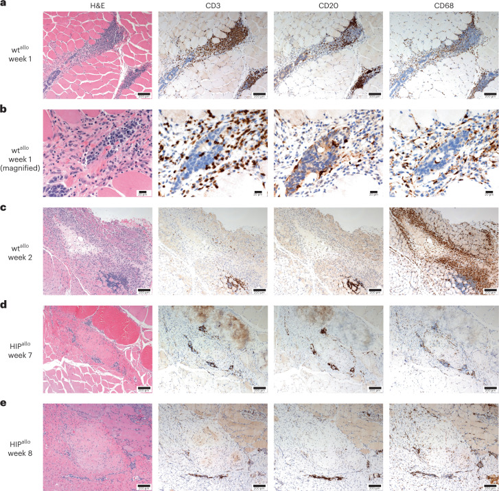
Fig. 4. Histology of intramuscular allogeneic rhesus macaque wt and HIP grafts.
Representative images of at least two independent sections are shown from H&E staining and immunohistochemical staining for CD3 (T lymphocytes), CD20 (B lymphocytes) and CD68 (macrophages). a,b, wtallo injection site after 1 week at ×100 (a) and ×400 (b) magnification. c, wtallo injection site after 2 weeks at ×100 magnification. d, HIPallo cell injection site after 7 weeks at ×100 magnification. e, HIPallo cell injection site after 8 weeks at ×100 magnification.

