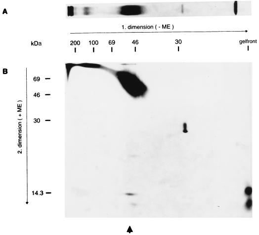
FIG. 6.
Radioimmunoprecipitation and two-dimensional electrophoresis. (A) The lysate of purified PrV(Ka) virions labelled with Tran35S-label (ICN) was precipitated with anti-gN serum. The precipitate was separated by SDS–10% PAGE under nonreducing (−ME) conditions and visualized by fluorography. (B) An identical lane was cut off the gel and placed horizontally on an SDS–13% acrylamide gel after incubation in reducing buffer (+ME). After electrophoresis, proteins were visualized by fluorography. The positions of molecular mass markers are indicated. The arrow denotes the position of the dissociated complex. ME, 2-mercaptoethanol.

