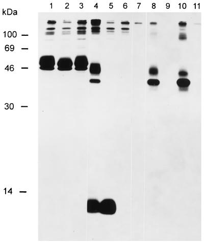
FIG. 7.
Demonstration of a gM-gN complex. Lysates of cells infected with PrV(Ka) (lanes 1, 4, and 8), PrV-ΔgMβ (lanes 2, 5, and 9), or PrV-gNβ (lanes 3, 6, and 10) or of noninfected cells (lanes 7 and 11) labelled with Tran35S-label (ICN) were precipitated with anti-gD serum (lanes 1 through 3), anti-gN serum (lanes 4 through 7), and anti-gM serum (lanes 8 through 11). Precipitates were separated by SDS–13% PAGE and visualized by fluorography. The positions of molecular mass markers are indicated on the left.

