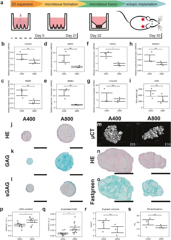
Figure 4.

Biological characterization of microtissues and ectopic explants. (a) Protocol overview and timeline. (b-g) mRNA quantification of differentiation markers. (i) DNA quantification on day 21. Histological staining of microtissues on day 21 showing (j) hematoxylin-eosin, (k) Alcian blue, and (l) safranin O. Scale bars = 200 μm. (m) Mineralisation after 4 weeks ectopic implantation of microtissue constructs measured by microCT. Histological staining of explants showing (n) hematoxylin-eosin, and (o) safraninO-fastgreen staining of representative explants. Scale bars = 1 mm. (p) Glycosaminoglycan (GAG) quantification. (q) Sulphated glycosaminoglycan (sGAG) quantification. (r) Explant volumes. (s) Explant percentage mineralisation.
