Figure 1.
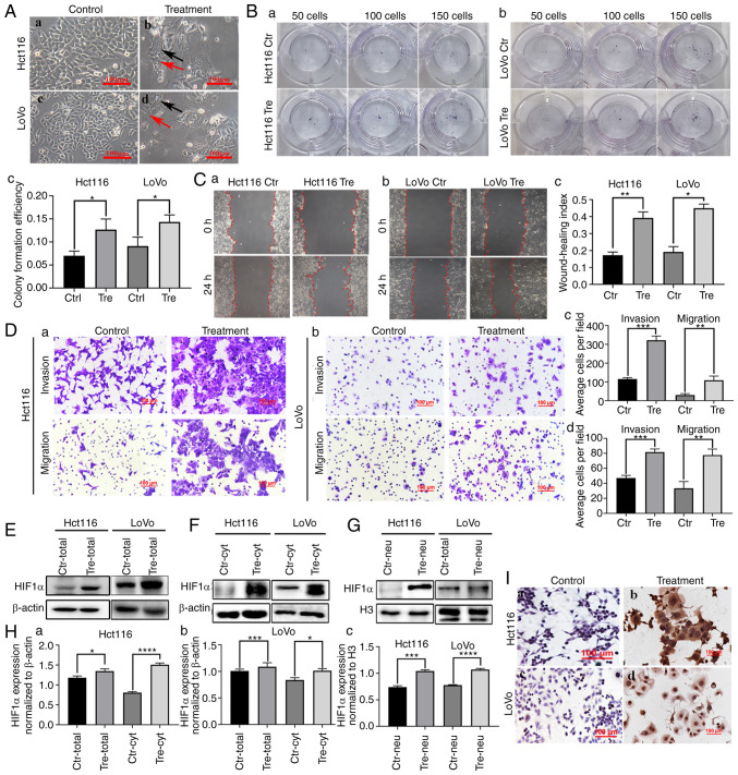
HIF1α expression in Hct116 and LoVo cells before and after CoCl2 treatment. (A) Control cells and PDCs derived from Hct116 and LoVo (magnification, ×100). (a) Hct116 control cells. (b) Hct116 PGCCs and daughter cells. (c) LoVo control cells. (d) LoVo PGCCs with daughter cells. The black arrow indicates PGCCs; the red arrow indicates PDCs. (B) Colony formation of 50, 100 and 150 (a) Hct116 control cells and PDCs, (b) LoVo control cells and PDCs and (c) statistiacal analysis of Colony formation efficiency of Hct116 and LoVo cells before and after CoCl2 treatment. (C) Wound healing assay of (a) Hct116 control cells and (b) LoVo control cells and PDCs at 0 h and 24 h (magnification, ×100). (c) Statistical analysis of wound healing index of Hct116 and LoVo cells before and after CoCl2 treatment. (D) The invasion and migration abilities of (a) Hct116 control cells and PDCs and (b) LoVo control cells and PDCs. Comparison of the average cell number in invasion and migration assay of (c) Hct116 PDCs and (d) LoVo PDCs before and after CoCl2 treatment. (E) Total HIF1α expression in Hct116 and LoVo control cells and PDCs. (F) Cytoplasmic and (G) nuclear HIF1α expression in Hct116 and LoVo control cells and PDCs. (H) Statistical analysis of total and cytoplasmic HIF1α expression in (a) Hct116 and (b) LoVo control cells and PDCs and (c) nuclear HIF1α expression in Hct116 and LoVo control cells. (I) Immunocytochemical staining of HIF1α in (a) Hct116 control cells, (b) Hct116 PDCs, (c) LoVo control cells and (d) LoVo PDCs. *P<0.05, **P<0.01, ***P<0.001, ****P<0.0001. HIF1α, hypoxia inducible factor 1 alpha; PDCs, daughter cells; PGCCs, polyploid giant cells; Tre, PGCCs with PDCs; Ctr, control.

