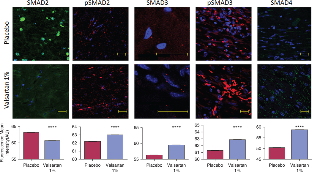
Figure 4. Changes in the transforming growth factor-beta superfamily downstream signaling proteins in wounds of aged diabetic pigs.
Valsartan-treated wounds express higher levels of SMAD3, phosphorylated SMAD2 and phosphorylated SMAD3, and the common mediator SMAD4 in healed skin compared with placebo. A decrease in the expression of SMAD2 was also observed in the valsartan-treated wounds. The photomicrographs present red or green fluorescent staining with a blue DAPI nuclear counter stain at ×63 magnification. Quantification of the levels of SMADs in porcine wounds is shown. Total n = 12 wounds per treatment group. Scale bar, 20 μm. The data are presented as the mean fluorescence intensity ± standard error of the mean. ****P < 0.0001. SMAD, mothers against decapentaplegic.

