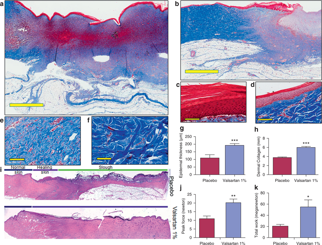
Figure 6. Impact of topical valsartan 1% on the quality of wound repair.

Masson’s trichrome staining of skin sections from aged diabetic pigs of (a) valsartan- and (b) placebo-treated wounds (scale bar, 4 mm). Representative image of epidermis in (c) valsartan- and (d) placebo-treated skin (scale bar, 50 μm). Representative image showing collagen arrangement in (e) valsartan- and (f) placebo-treated skin (scale bar, 50 μm). (g, h) Quantification of thickness of epidermis and dermal collagen layers in porcine wounds. (i) Hematoxylin and eosin image. Biomechanical assessment of healed skin in pig cohorts showing comparison of the (j) average tension at the break point and (k) average work at the break point in healed wounds. Total n = 12 wounds per treatment group. *P < 0.05, **P < 0.01, ***P < 0.001.
