
FIGURE 7.
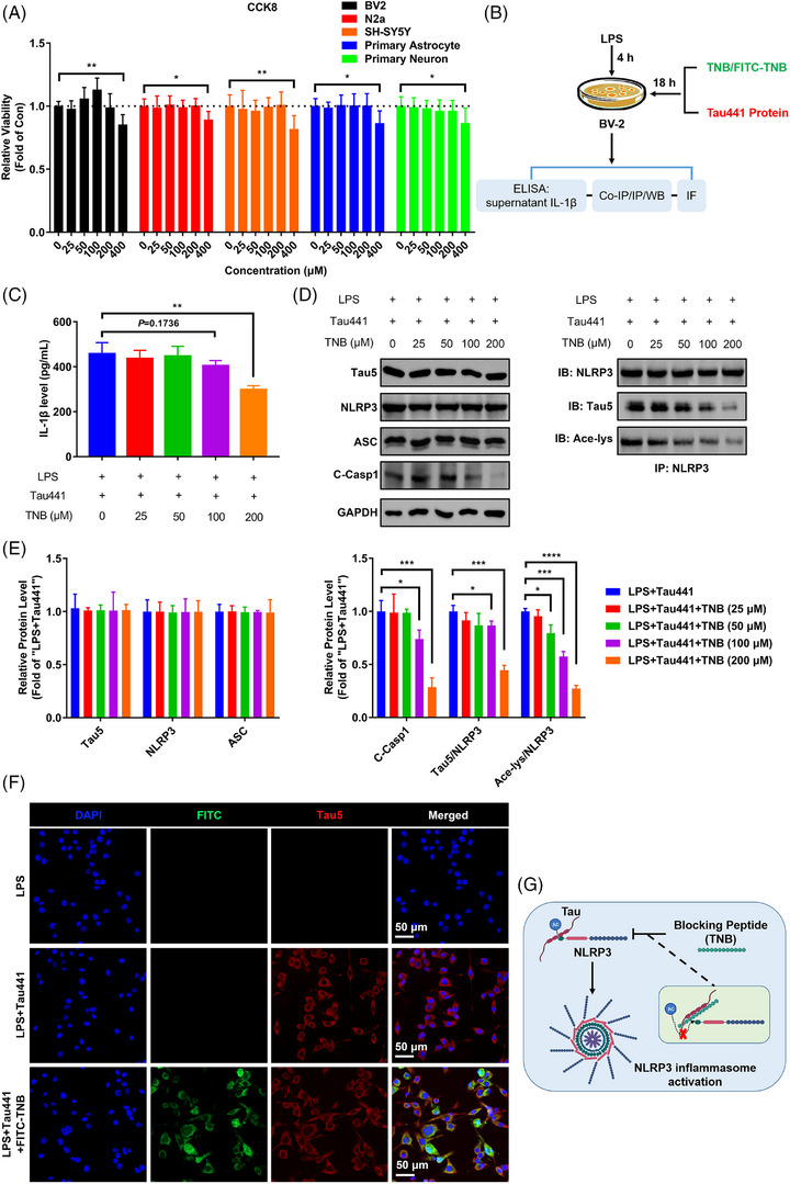
Tau–NLRP3‐binding blocking (TNB) peptide blocks the binding of Tau to nucleotide‐binding oligomerisation domain (NOD)‐like receptor pyrin domain containing 3 (NLRP3), prevents NLRP3 acetylation and inflammasome activation in BV‐2 cells. (A) CCK8 detection for the viability of different cells (BV‐2, N2a, SH‐SY5Y, primary astrocyte and primary neuron) after TNB peptide treatment at different dosages (0, 25, 50, 100, 200 and 400 µM) for 48 h. n = 6, * p < .05, ** p < .01 as indicated. (B) Schematic diagram of in vitro experiments for testing TNB blocking function and downstream effect. (C) Enzyme‐linked immunosorbent assay (ELISA) for detecting interleukin‐1β (IL‐1β) production of LPS and Tau441 protein challenged BV‐2 microglia with or without TNB peptide treatment at different dosages (0, 25, 50, 100 and 200 µM). n = 3, ** p < .01 as indicated. (D) The ability of TNB peptide of blocking Tau–NLRP3 binding and its effects on Tau‐induced NLRP3 acetylation and inflammasome activation were measured by immunoprecipitation and immunoblotting using anti‐Tau5, anti‐NLRP3, anti‐ASC, anti‐cleaved Caspase‐1 (C‐Casp1), anti‐GAPDH and anti‐acetylated‐lysine (Ace‐lys) antibody. (E) Quantification of the blots in (D). The ratio of Tau5 to NLRP3 stands for the Tau–NLRP3‐binding ability (immunoprecipitated Tau by anti‐NLRP3/NLRP3). The ratio of Ace‐lys to NLRP3 stands for the level of NLRP3 acetylation. n = 3, * p < .05, *** p < .001, **** p < .0001 as indicated. (F) Representative images of exogenous human Tau441 protein immunofluorescence (anti‐Tau5, red) with the nuclei and TNB peptide labelled by DAPI (blue) and FITC (green) respectively, showing the co‐localisation of TNB peptide and Tau441 protein in BV‐2 cells. Scale bar = 50 µm. (G) Mechanism diagram showing TNB peptide intercepts the binding of Tau to NLRP3, thereby inhibiting NLRP3 acetylation and preventing inflammasome activation.