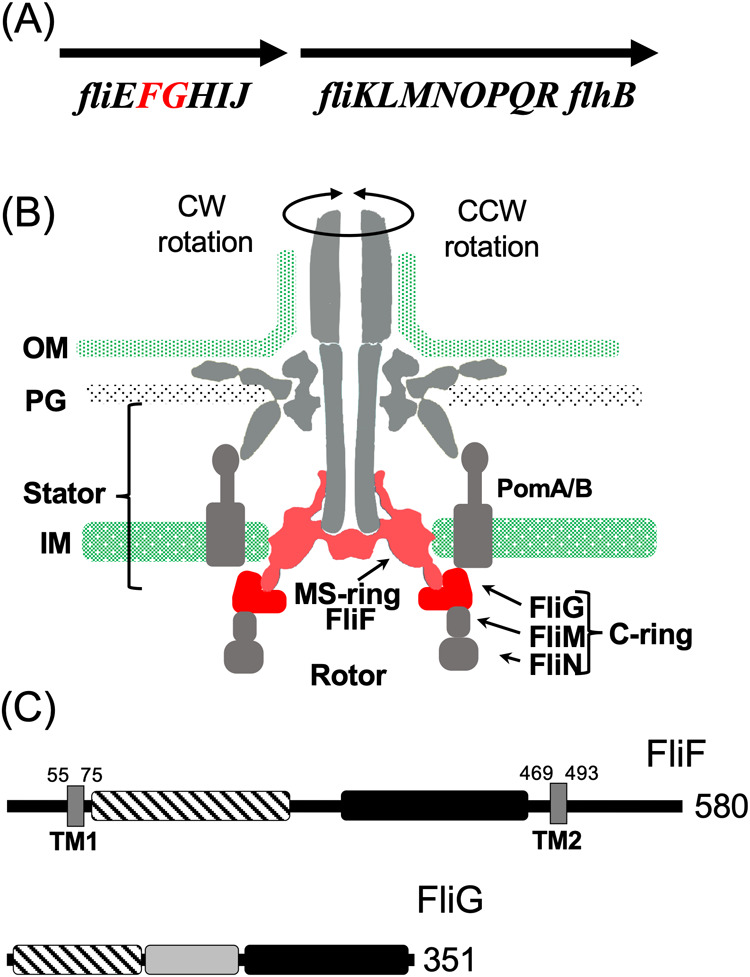
Figure 1 .

Flagellar motor structure and the component genes. (A) The genomic context of the flagellar genes around fliF and fliG. (B) The longitudinal section image of the flagellar motor. The C-ring is composed of FliG, FliM, and FliN. The MS-ring is composed of FliF. The stator is composed of PomA and PomB. FliF and FliG are indicated in red. (C) The primary structure of FliF and FliG. The distinctive regions of the protein are represented by the squares painted with different patterns. The dark box, stripe box, and filled box indicate the transmembrane (TM) region, M-ring region and S-ring region, respectively, for FliF. The stripe box, dark box, and filled box indicate the N-terminal region, the middle region, and C-terminal region, respectively, for FliG. The numbers indicate the amino acid residues.
