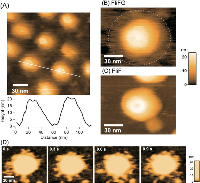
Figure 5 .

High-speed atomic force microscopy (HS-AFM) images of the purified Vibrio MS-ring. (A) HS-AFM image of MS-ring comprising FliFG fusion proteins and the cross-sectional profile along the white line on the image. (B) Magnified image of MS-ring composed of the FliFG fusion proteins. (C) HS-AFM image of MS-ring composed of the FliF proteins. (D) Clipped images of the MS-ring composed of FliFG fusion proteins with enhanced contrast of the lower part. Imaging rate: 0.15 s/frame.
