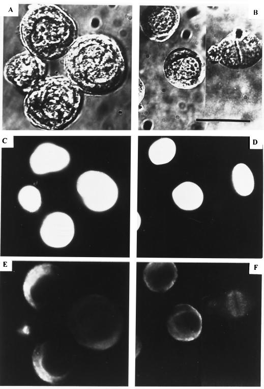
FIG. 1.
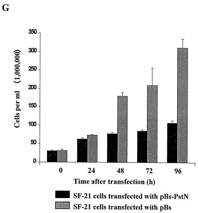
Expression of the AcMNPV PstI-N fragment blocks cell division. SF-21 cells (0.5 × 10⁶ per 35-mm-diameter dish) were transfected either with 0.5 μg of plasmid pBs-PstN (A, C, and E) or with the same amount of the control vector plasmid, pBs (B, D, and F). At 48 h posttransfection, the cells were fixed in methanol and then double stained with DAPI (C and D) and DM1 anti-alpha tubulin (E and F), and the same field of cells was examined by phase-contrast (A and B) and immunofluorescence (C to F) microscopy. All cells were photographed at the same magnification; note the larger sizes of the cells transfected with pBs-PstN. Also, one of the four cells in panels A, C, and E does not appear to have been transfected with pBs-PstN. In panels B and F, mitotic spindles can be observed in the cell on the right. Bar, 25 μm. (G) SF-21 cells were transfected with either pBs or pBs-PstN and harvested after 24, 48, 72, and 96 h, and the numbers of viable cells were counted in the presence of trypan blue.


