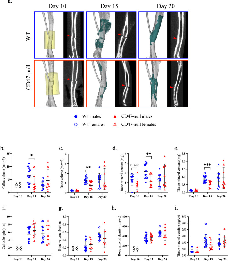
Figure 6. Genetic knockout of CD47 inhibits early ischemic fracture callus formation.
µCT analysis of ischemic tibia fracture callus of WT (n=7–11) and CD47-null (n=6–9) mice at day 10, 15, and 20 post-fracture. a, Representative 3D reconstructions (white background) and sagittal-plane reconstruction (black background) of WT (top row) and CD47-null (bottom row) mice at day 10, 15, and 20 post-fracture. Location of fracture (red arrowhead) is marked on the sagittal reconstructions. Day 10 3D reconstruction includes representative cylindrical ROIs (transparent yellow cylinder) used to calculate callus morphology. Day 15 and 20 representative 3D reconstruction include highlighted callus mineralization (teal). b-i, Callus morphology (mean±SD) at days 10, 15, and 20 post-fracture. *P<0.05, **P<0.01, ***P<0.001, two-sided t tests performed at each timepoint; § no data.

