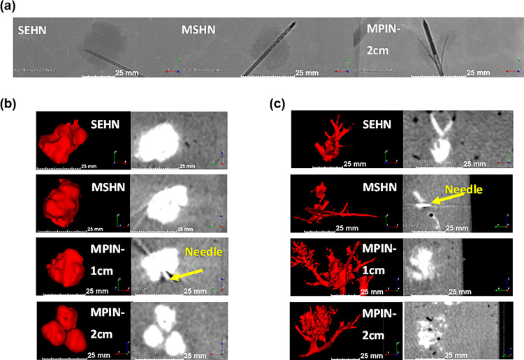
Figure 3.
Fluoroscopic and cone beam CT (CBCT) imaging following injection of 4 mL of POL22 with iodine or iodine alone at 10 mL/hr into ex vivo bovine liver. Three needles were used: SEHN, MSHN, and MPIN deployed at 1 cm (MPIN-1 cm) and deployed at 2 cm (MPIN-2cm). (a) Fluoroscopic images following injection of POL22 with iodine with SEHN, MSHN and MPIN-2cm showing the gel deposition (higher density areas) relative to the needle locations. (b) POL22 with iodine injections. 3D surface renderings (left) and corresponding cross-sectional images (right) based on CBCT imaging. (c) Injections of iodine alone. 3D surface renderings (left) and corresponding cross-sectional images (right) based on CBCT imaging

