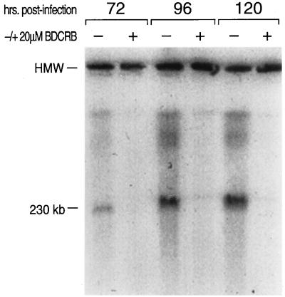
FIG. 3.
Inhibition of HCMV DNA concatemer processing by BDCRB. HCMV-infected cells were grown with and without BDCRB added immediately following infection. HMW DNA was separated by CHEF gel electrophoresis and located in the gel by Southern analysis with 32P-radiolabelled HCMV DNA fragments. The image was captured by exposure of the filter to a PhosphorImager screen and analyzed with ImageQuant software. The band at the top of the gel, labelled HMW, is the well where most of the DNA remained. The 230-kb band is HCMV DNA migrating at the position expected for genome-length DNA. Concatemeric phage lambda genomic DNA was run on the same gel to serve as molecular size standards.

