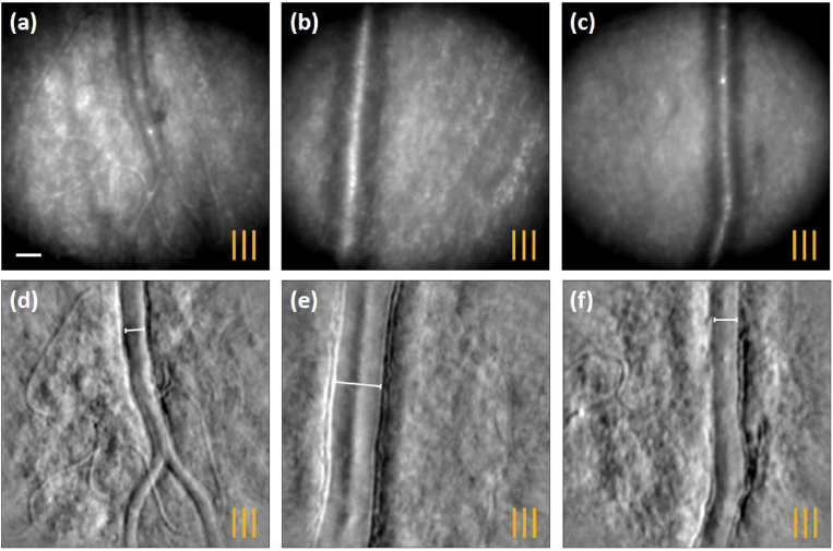
Fig. 8.
Registered average of confocal retinal vessel images of (a) N1 at T SR, (b) N2 at T IR, (c) N3 at T SR obtained with vertical line illumination. (d)-(f) The corresponding phase contrast images acquired simultaneously. The orange lines indicate the orientation of the line illumination. The scale bar is 20 m.

