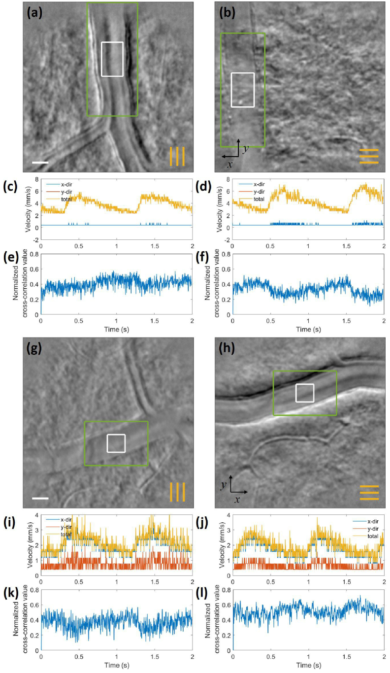
Fig. 10.
Registered average of phase contrast vascular images of N2 at T SR using (a) vertical line illumination and (b) horizontal line illumination. (c)-(d) show the velocity measurement over 2 seconds showing average velocity of 4 mm/s. (e)-(f) Corresponding normalized cross-correlation values, showing inverse relationship with the flow velocities. (g)-(h) show similar images of N3 at T IR. (i)-(j) Blood flow velocity measurement from (g)-(h) showing average velocity of 2 mm/s. (k)-(l) Corresponding normalized cross-correlation values, showing lower normalized cross-correlation values with illumination lines perpendicular to the vessel than those parallel to the vessel. The orange lines indicate the orientation of the line illumination. The scale bar is 20 m.

