. 2024 Jan 5;12:RP90133. doi: 10.7554/eLife.90133

Figure 2. Bsh activates Ap/Pdm3 expression and specifies L4/L5 neuronal fate.

(A–B’’) Bsh-knockdown (KD) in lamina progenitor cells (LPCs) (R27G05-Gal4>UAS-Bsh-RNAi) eliminates Bsh in LPCs and neurons in wandering L3 (white circle). Tll labels all LPCs. LF: lamina furrow. L3: third larval instar. Here and below, scale bar: 10 µm, n≥5 brains. (C) Schematic of lamina neuron development at third larval instar. (D) Schematic of lamina neuron development from 1-day pupae to adult. (E–F’’) Bsh remains undetectable in the lamina of 3- to 4-day pupa in Bsh-KD (R27G05-Gal4>UAS-Bsh-RNAi). (G–K) Bsh-KD in LPCs removes most L4 (Bsh/Ap) (J) and L5 (Bsh/Pdm3) (I) neuron markers. The Ap expression in L5 is caused by the Gal4 driver line but is irrelevant here. (K) The number of Elav+ cells in a single slice. n=5 brains in (I) and (J), n=7 brains in (K). L4 layer, yellow outline. L5 layer, white outline. (L) Summary. Data are presented as mean ± SEM. Each dot represents a brain. ***p<0.001, ns = not significant, unpaired t-test.

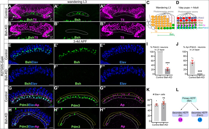
Figure 2.

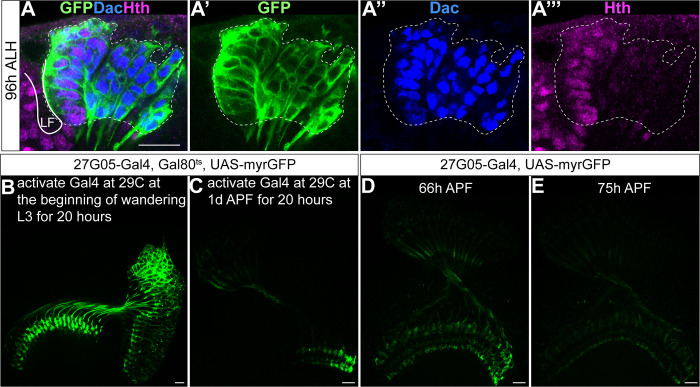
Figure 2—figure supplement 1. R27G05-GAL4 is turned on in all lamina progenitor cells (LPCs) and turned off in lamina neurons.

Figure 2—figure supplement 1.

(A–A’’’) R27G05-Gal4 drives GFP expression in all LPCs and lamina neurons (white dash circle) which are labeled by Dac at 96 hr after larval hatching (ALH). Hth labels LPCs next to lamina furrow (Piñeiro et al., 2014). Here and below, scale bar, 10 µm. (B, C) R27G05-Gal4 is turned off in lamina neurons. Activating R27G05-Gal4 at 29C at the beginning of wandering L3 for 20 hr (by inactivating Gal80ts) reveals continued expression of GFP; in contrast, activating R27G05-Gal4 at 29C at 1 day after pupa formation (APF) for 20 hr reveals little GFP, showing that R27G05 is not expressed in pupal stages containing postmitotic lamina neurons. Genotype: R27G05-Gal4>UAS-myrGFP, tubP-Gal80[ts]. (D, E) Although R27G05-Gal4 is turned off by 1 day APF, GFP persists in lamina neuron cell bodies until 66 hr APF and in their neurites until 75 hr APF. Genotype: R27G05-Gal4>UAS-myrGFP.
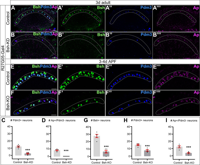
Figure 2—figure supplement 2. Bsh-knockdown (KD) or Bsh-knockout (KO) in lamina progenitor cells (LPCs) affects L4/L5 neuronal fate.

Figure 2—figure supplement 2.

(A–D) Bsh-KD in LPCs removes most L4 (Bsh/Ap) and L5 (Bsh/Pdm3) neuron markers of 3-day adult flies. The Ap expression in L5 is caused by the Gal4 driver line but is irrelevant here. (C and D) The number of Pdm3+ (C) or Ap+/Pdm3- (D) cell bodies in a single slice in control (A) and Bsh-KD (B). Here and below, scale bar, 10 µm, white circle outlines the region of L4 and L5 cell bodies. (E–I) Bsh-KO in LPCs (27G05-Gal4>UAS-Cas9, UAS-Bsh-sgRNAs) decreases Ap and Pdm3 expression in the lamina of 3- to 4-day-old pupa. (G–I) The number of Bsh+ (G), Pdm3+ (H), or Ap+/Pdm3- (I) cell bodies in a single slice in control (E) and Bsh-KO (F). Data are presented as mean ± SEM. Each dot represents each brain. n=6 brains in (C) and (D), n=5 brains in (G), (H), and (I). Each dot represents each brain. ***p<0.001, unpaired t-test.
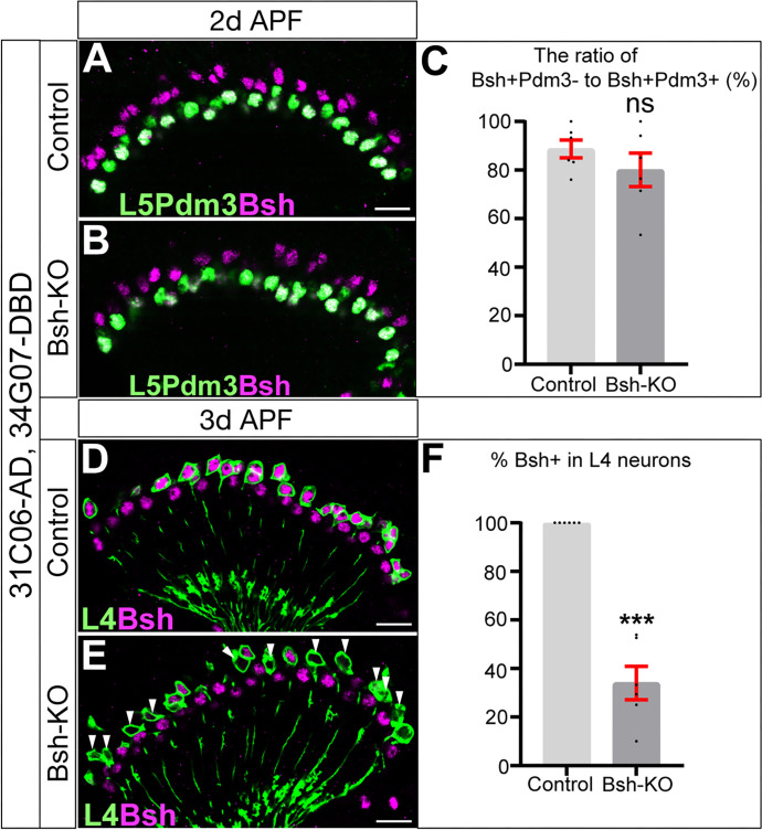
Figure 2—figure supplement 3. Bsh-knockdown (KD) in L4 neurons results in loss of detectable Bsh between 2 and 3 days after pupa formation (APF).

Figure 2—figure supplement 3.

(A–C) Bsh expression is detected at 2 day APF following Bsh-knockout (KO) in L4 neurons. Genotype: 31C06-AD, 34G07-DBD>UAS-Cas9, UAS-Bsh-sgRNAs. (D–F) Bsh expression is absent at 3 days APF following Bsh-KD in L4 neurons. Genotype: 31C06-AD, 34G07-DBD>UAS-Cas9, UAS-Bsh-sgRNAs. GFP labels L4 neurons. White arrowhead indicates L4 cell body lacking Bsh expression. Data are presented as mean ± SEM. ns = not significant, ***p<0.001, unpaired t-test, n=6 brains; each dot represents one brain. Scale bar, 10 µm.
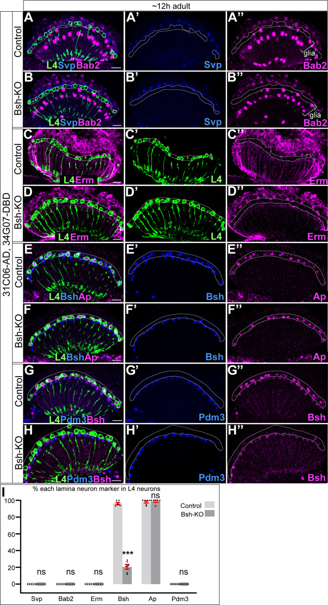
Figure 2—figure supplement 4. Bsh is not required in L4 neurons to maintain L4 neuronal fate.

Figure 2—figure supplement 4.

(A–B’’) L1 marker Svp and L2 marker Bab2 are not detected in L4 cell bodies when knocking out Bsh in L4 (31C06-AD, 34G07-DBD>UAS-Cas9, UAS-Bsh-sgRNAs). Here and below, L4 is labeled by GFP (31C06-AD, 34G07-DBD>UAS-myrGFP). White outline, L4 cell body. Scale bar, 10 µm. (C–D’’) L3 marker Erm is not detected in L4 cell bodies when knocking out Bsh in L4. (E–F’’) Bsh becomes absent in most L4 while Ap expression remains normal when knocking out Bsh in L4. (G–H’’) L5 marker Pdm3 is not detected in L4 cell bodies when knocking out Bsh in L4. (I) The percentage of each lamina neuron marker in L4 neurons (GFP+). ***p<0.001, ns = not significant, unpaired t-test, n=5 or 6 brains, each dot represents each brain, data are presented as mean ± SEM.