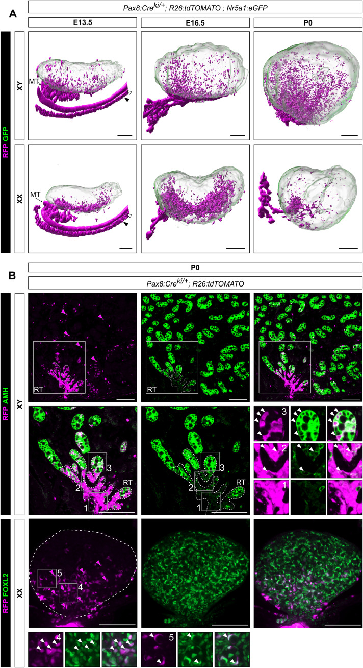
Fig. 5. Pax8+ progenitors contribute to both rete testis/ovarii and the pool of Sertoli and pregranulosa cells.
(A) 3D reconstructions of Pax8:Cre;Rosa26:tdTomato;Nr5a1:GFP testis and ovary at E13.5, E16.5, and P0. Pax8+ cells are lineage-traced with RFP. Green fluorescent protein (GFP), expressed under the control of the Nr5a1 promoter, is used to delineate gonadal cells. Note the presence of RFP+ cells close to the rete testis/ovarii but also throughout the gonads. White arrowhead indicates the Wolffian duct, and black arrowhead the Müllerian duct. (B) Double IF for RFP/AMH and RFP/FOXL2 respectively in XY and XX Pax8:Cre;Rosa26:tdTomato;Nr5a1:GFP mice at P0. Note the presence of RFP+ cells increases near the rete testis and rete ovarii. Cells in the rete testis are exclusively RFP+ and do not express AMH (inset 1). At the junction between the rete testis and the testis cords, low AMH expression is observed in some RFP+ cells (inset 2). RFP+/AMH+ cells are also present in testis cords (inset 3). Similarly, RFP+/FOXL2+ cells are present in pregranulosa cells of the developing ovary (inset 4), including in some primordial follicles (inset 5). Magenta arrows indicate RFP+ cells, and white arrows indicate RFP+/AMH+ or RFP+/FOXL2+ cells. MT, mesonephric tubules; RT, rete testis. Scale bars, 200 μm in (A) and 100 μm in (B).

