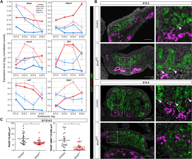
Fig. 6. Wnt4 is required for rete testis formation.
(A) Expression profiles of genes involved in the WNT/β-catenin pathway in SLCs and supporting cells. Data were extracted from scRNA-seq analysis of XX and XY embryos at E11.5, E12.5, E13.5, and E16.5. (B) Representative double IF against PAX8 and AMH in control (Sf1:Cre;Sox9F/+;Wnt4KO+/+) or Wnt4KO (Sf1:Cre;Sox9F/+;Wnt4:KO−/−) XY embryos at E12.5 and E14.5. Boxes indicate regions shown on the right magnifying the rete testis and adjacent testis cords. Note that the numbers of PAX8+ cells, the rete testis, and the testis cords near the rete testis are reduced and disorganized in Wnt4 mutant embryos both at E12.5 and E14.5. White and yellow arrowheads indicate PAX8+ and PAX8+/AMH+ cells, respectively. DAPI was used as a nuclear counterstain. Scale bars, 100 μm. (C) Quantification of PAX8+ and PAX8+/AMH+ cells per 10,000 μm2 of testis section of control and Wnt4KO embryos at E14.5. Data are presented as a box-and-whisker plot to illustrate the heterogeneity of PAX8-positive cells according to the different sections. Each point represents the quantification of one testicular section. Three embryos per genotype were used. Student’s t test, two-sided unpaired (*P < 0.05).

