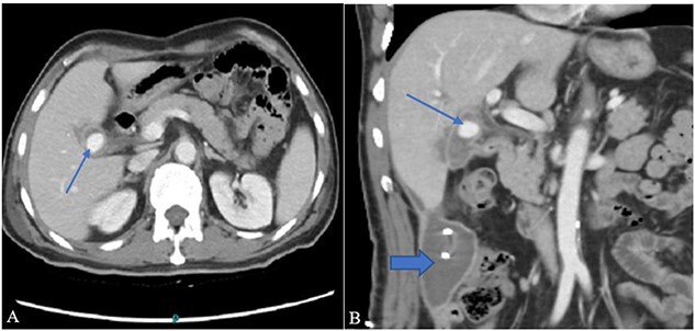
Figure 1.

(A) Contrast-enhanced CT axial image through the abdomen shows a small walled-off collection in the GB fossa with a central contrast density representing pseudoaneurysm (arrow). The collection appears to be in communication with the second part of the duodenum. (B) A coronal image from the same CT shows a right para colic gutter collection with an in situ pigtail (broad arrow).
