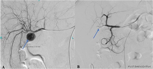
Figure 2.

(A) Pre-embolization angiogram of the hepatic artery demonstrates pseudoaneurysm of the cystic artery stump (arrow). (B) Post-coil embolization angiogram of the hepatic artery shows no further filling of the pseudoaneurysm (arrow).

(A) Pre-embolization angiogram of the hepatic artery demonstrates pseudoaneurysm of the cystic artery stump (arrow). (B) Post-coil embolization angiogram of the hepatic artery shows no further filling of the pseudoaneurysm (arrow).