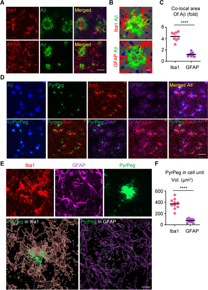
Fig. 1.
Microglia that contain aggregated amyloid-β are activated and are located near neuritic plaques in 5XFAD mice. A Z-stack images of brain tissue from 5XFAD mice after staining with Iba1 or GFAP in the peri-plaque region. Scale bar: 200 µm. B Three-dimensional reconstruction of Z-stack images in which Aβ overlaps with staining by anti-Iba1 antibody or anti-GFAP antibody. Scale bar: 10 µm. C Quantification of Aβ volumes co-localized with glial volumes, as measured in cell units. ****p < 0.001, Aβ volume in Iba1 volume versus Aβ volume in GFAP volume; unpaired Student’s t test. D Representative images of 5XFAD mouse brain sections stained with an anti-Aβ antibody (blue), PyrPeg (green), anti-Iba1 antibody (red), and anti-GFAP antibody (magenta). Scale bar: 100 µm. E Orthogonal view of a 3-dimensional reconstruction of confocal images showing co-localization of an Aβ plaque (green) with Iba1(pink) or GFAP (purple). Scale bar: 10 µm. F Quantification of PyrPeg co-localized with microglia versus astrocytes, as measured in cell units. ****p < 0.001, PyrPeg volume in Iba1 volume versus PyrPeg volume in GFAP volume; unpaired Student’s t test

