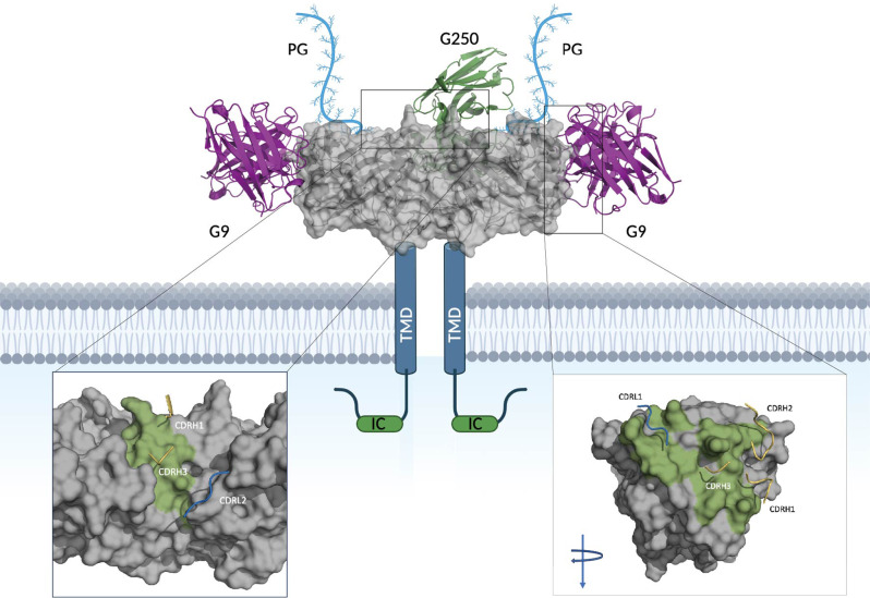
Fig. 6.
Binding mode of G9 and G250 with CAIX. Depicts TAA, CAIX, gray, along with its transmembrane domain, intracellular tail and proteoglycan (PG) domain at the N-terminus. The variable regions of mAb, G9, are shown in pink bound to the predicted epitope and the variable regions of mAb G250, are shown in green. The zoomed view depicts the binding interface of G250 to CAIX (left) and G9 to CAIX (right, turned 90 degree) with epitopes colored in green and contact regions of the complementarity determining regions (CDRs) shown in yellow (heavy chain), and blue (light chains). G9 shows 2:1 binding mode to a CAIX dimer and G250 binds to a CAIX dimer in 1:1 ratio

